
- français
- English
2015/report/debugging/box
[EXPLIQUER LA BOITE, EXPLIQUER NOS RESULTATS, AJOUTER DES PASSAGES DE LA BIODESIGN ENTRY]
We are using spectruino to quantify how much light scattering influences the detection of green fluorescence. We would like to find if the blue light, which excites GFP in bacteria, is also detected by the sensor because it can’t distinguish between green and blue light. We tested several samples : Free GFP from cell Lysis, eGFP within bacteria, fluorescent dextran and cell lysed without GFP (control), in order to compare the spectra and observe scattering.
So we tried to test with the prototype but we noticed the length was too big and the spectruino didn’t detect any spectrum. So we decided to build a smaller box
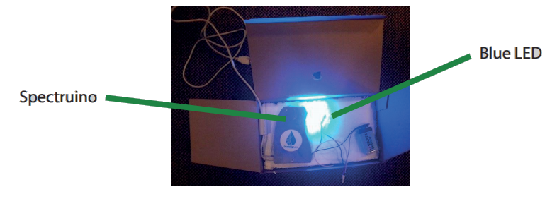
The blue LED goes through the vial and spectruino detects the resulting spectrum. The spectruino is connected to a computer which displays the spectrum.
We chose a closed box in order to eliminate the stray light. The second step was to fix the vial, the blue LED and the spectruino with the right angle, in order to have the best detection of the spectrum. We used polystyrene to stabilize the system because it can be found anywhere for free. And we added a hole to facilitate the changing of the vial.
Now that we have the samples AND the test box, we can finally quantify the light scattering. We obtained different spectra with our 4 samples
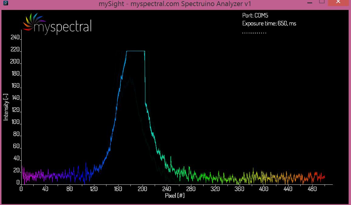
eGFP bacteria sample
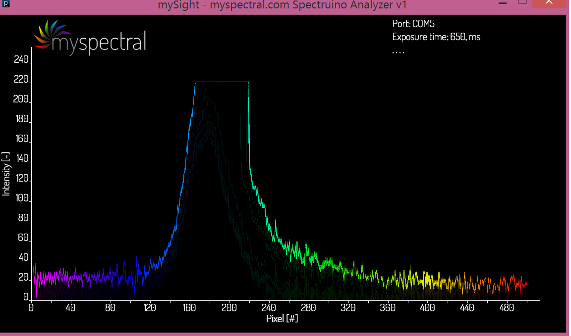
We observe one big blue peak which is a little bit extended on the green spectrum
Free GFP sample:
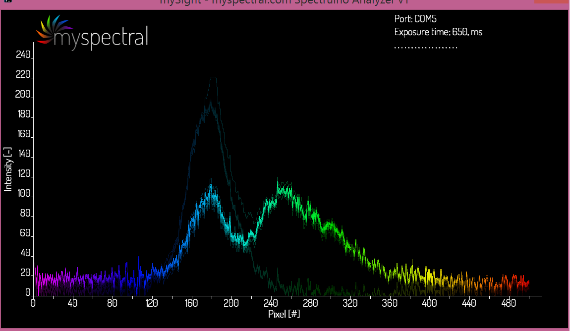
We observe two peaks: one on blue spectrum, one on green spectrum. The two peaks have the same amplitude.
Fluorescent dextran:
We observe two peaks: one on bigger blue spectrum, one on smaller green spectrum
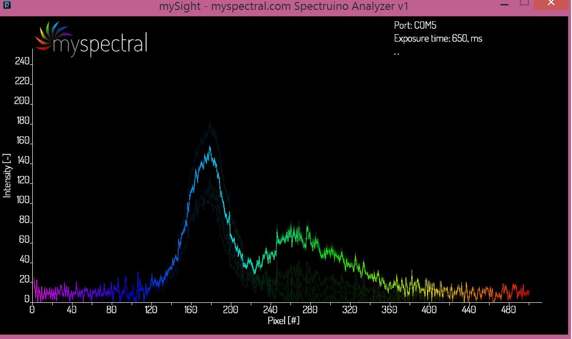
lysed cell without GFP (control):
We observe one blue peak
Our analysis:
We would like to observe two peaks in eGFP bacteria sample, because it would mean that the scattering isn’t important. But we observe only one peak and the green peak is hidden by the big blue peak.
We estimate that the blue peak in Control sample has a smaller intensity than eGFP bacteria sample. That means the bacteria scattered the blue light and therefore the spectruino detected more blue light that there is in reality.
We obtained two peaks with the dextran sample, as well as with the free GFP sample. We know that the dextran does not scatter so we can conclude that free GFP does not scatter as well. The bigger blue peak in dextran sample can be explained by the structural difference between dextran, which is a sugar polymer, and GFP. So the light does not go through the same way.
To conclude, the free GFP sample gives two distinct peaks, which demonstrate that there is no scattering anymore. There is more green fluorescent and less blue light without scattering and so the detector would detect the right amount of green fluorescent without stray blue light. That is very satisfying! Cell lysis is a good way to prevent light scattering. Unfortunately cell lysis has to take place after GFP expression and so after the mixing with the water sample. We couldn’t send the lysed cell, so the people which receive the kit should lyse the cell.