
- français
- English
2015/report/debugging/cell-lysis
We drew our inspiration for the protocol from the following paper: « William W Ward of Brighter Ideas, Three-Phase Partitioning for Protein Purification ». We chose the Three-Phase Partitioning method because it seemed to be an easy and interesting method to separate GFP from bacteria. We were following the protocol from the paper but at the step where we should have seen if the concentration of the starting buffer was good we didn't obtain the good result and each concentration seemed to be higher. We decided to go our way and to try with three different concentrations (1.6 M, 3M and 4M) of starting buffer. Finally the result gave that the GFP was not visible to the naked eye but with fluorescence analysis the GFP was existing in the 3 samples. The 1.6M of starting buffer has the higher green fluorescence value so we decided to perform our protocol with this concentration. We performed again the experiment with more bacteria to obtain more green fluorescence with this following protocol:
MATERIAL:
- 1.6 M starting buffer composed of:
- 10.57g of ammonium sulphate
- 2.5 ml of 50nM of Tris Buffer pH 8
- add water until 50ml
- eGFP bacteria
- Ars reporter control bacteria
- tert-Butanol (ref: 19460)
PROTOCOL:
- Compare absorbance of eGFP bacteria and bacteria control (to see if we can put the same volume)
- Take 50ml of eGFP bateria and 50ml of control bacteria
- Centrifuge 2 tubes at 10 000 rpm(or the max on the bug centrifugeuse) during 10 min (to convert rpm in rcf RCF = 1.1118 x 10⁻⁵ x r x rpm^2)
- Remove supernatant, we only keep the pellet bacteria
- Add 5ml of 1.6M of starting buffer and 5ml of t Butanol in the 2 tubes
- Vigourous shaking during 1min (vortex)
- Centrifuge during 10min at 1000rpm
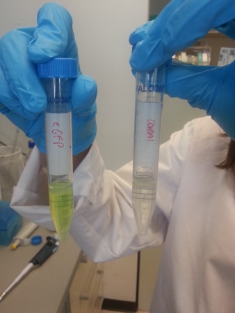
- Observe 3 phases: butanol, membrane and constituent cells, GFP phase for eGFP bacteria and without GFP for control.
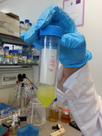
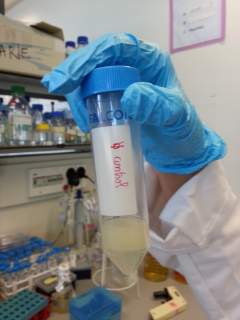
- Keep only the lowest phase, which is GFP phase for eGFP bacteria and without GFP for control and remove the rest
- Add 500μL of t Butanol
- Centrifuge 2 tubes at 2000rpm during 10min
- We obtained again 3 same phases
- Keep only the lowest phase, which is GFP phase for eGFP bacteria and without GFP for control and remove the rest
- Measure green fluorescence , excitation at 488nm


RESULTS:
absorbance of eGFP bacteria: 1.96 OD
absorbance of control bacteria: 2 OD
so we can put the same volume (50ml) because the OD values are close enough
fluorescence result:
eGFP: 45083
control bacteria: 873
1.6M starting buffer: 1
CONCLUSION:
We measured the fluorescence of 1.6M starting buffer to compare the green fluorescence between bacteria with GFP, bacteria and sample without bacteria.
The fluorescence of control bacteria of 873 can be explained because lots of things fluoresce in green in biology. The difference between eGFP and control bacteria fluorescence is significant.
We obtained about 4ml of free GFP (from lysis of eGFP bacteria).
REFERENCE: [1] William W Ward of Brighter Ideas, Three-Phase Partitioning for Protein Prufification [pdf]