
- français
- English
2015/report/debugging/intro
By reading the last final report (Report 2014), we saw that the prototype detected much more fluorescence in the most concentrated bioreporter solution than the lab equipment. We did the assumption that this problem could be due to light scattering of the blue LED because bacteria's shape might deviate this blue light. So, our part of the project was to quantify, observe and test the light scattering in the prototype.
To determine if the sensor also detects blue light, we wanted to compare eGFP bacteria and lysed eGFP bacteria. To test this assumption we realized a cell lysis protocol. We chose the Three-Phase Partitioning method inspired by "William W Ward of Brighter Ideas, Three-Phase Partitioning for Protein Prufification" [pdf]
Once the cell lysis protocol was performed, we analysed the light spectrum of different samples using spectruino to compare the spectrum of green fluorescense and blue light in eGFP bacteria and free GFP from lysed cell. The different samples were: Free GFP from cell lysis, eGFP bacteria, fluorescent dextran as negative control because we know that dextran does not scatter and fluorescent beads as positive control.
This figure shows what we would observe if the light scattering were present:
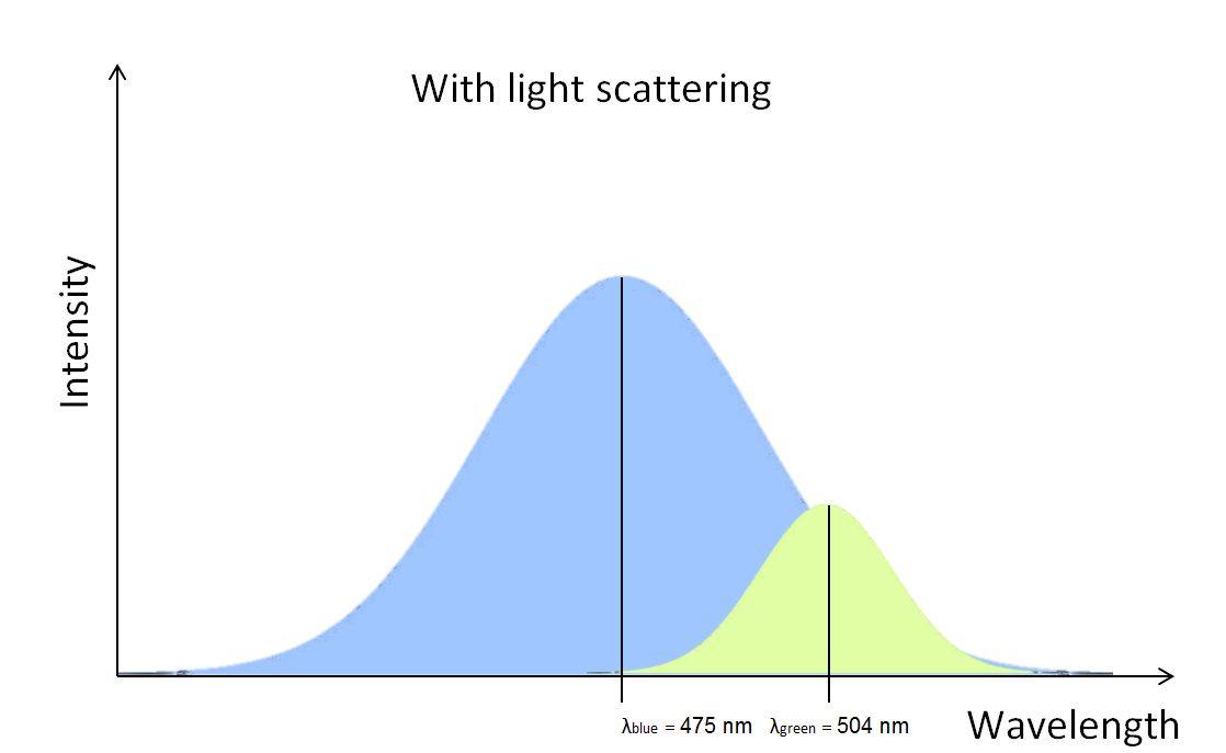
The green fluorescense disappears in the blue light.
This figure shows what we would observe if the light scattering were small:
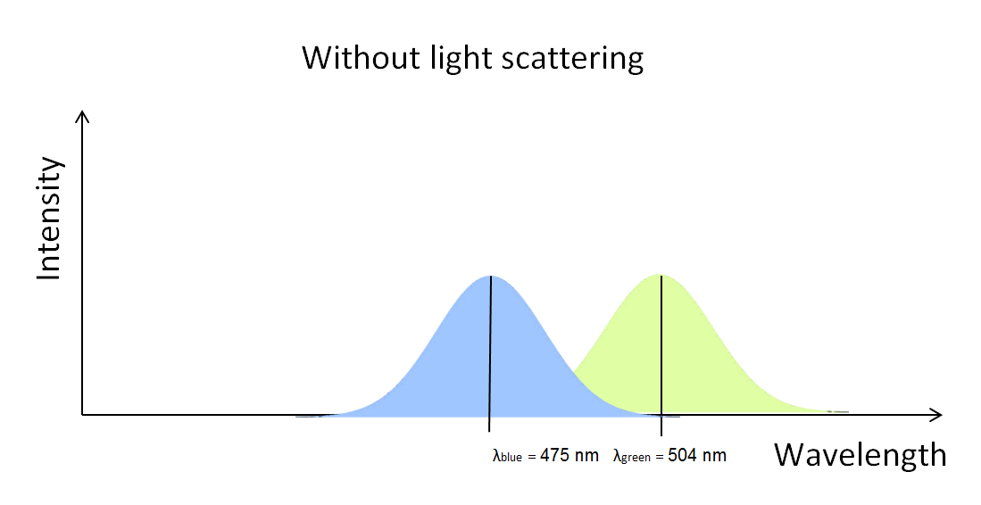
We would be able to observe two well separated peaks.
If the light scattering were significative, the eGFP bacteria spectrum would look like the first figure. And if cell lysis would decrease light scattering significatively, the free GFP from cell lysis spectrum would look like the second figure.
We tried to test directly with the prototype, after having exchanged the sensor by the spectruino, but unfortunately that didn’t work because the length between the spectruino and the vial was too high. To solve this problem, we built a box serving only to test the light scattering.